|
ICP811S - INTERGRATED CLINICAL PATHOLOGY - 1ST OPP - JUNE 2023 |
|
|
1 Page 1 |
▲back to top |
|
|
2 Page 2 |
▲back to top |
|
|
3 Page 3 |
▲back to top |
|
|
4 Page 4 |
▲back to top |
|
|
5 Page 5 |
▲back to top |
|
|
6 Page 6 |
▲back to top |
|
|
7 Page 7 |
▲back to top |
|
|
8 Page 8 |
▲back to top |
|
|
9 Page 9 |
▲back to top |
|
|
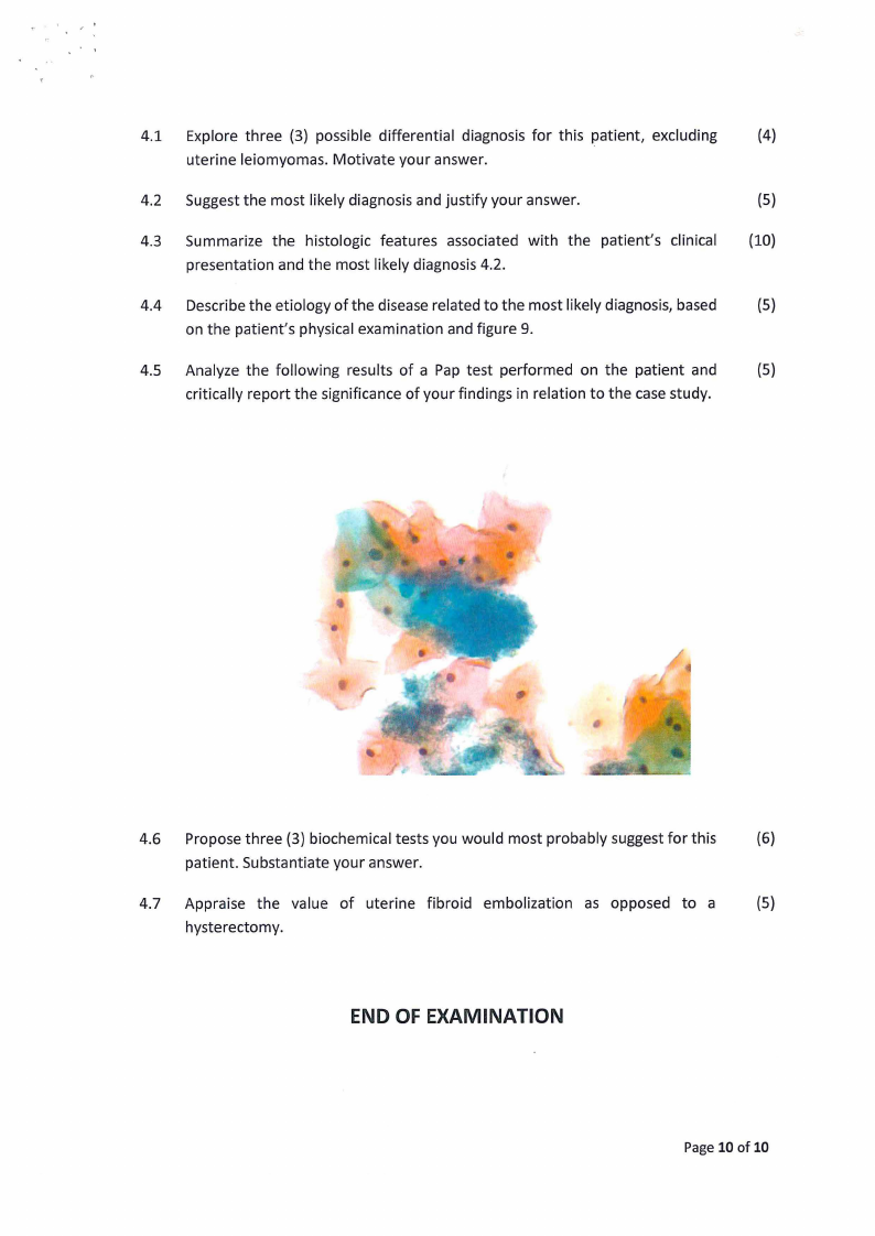
10 Page 10 |
▲back to top |