 |
ANP611S - ANATOMICAL PATHOLOGY 2A - 2ND OPP - JULY 2023 |
 |
1 Page 1 |
▲back to top |
nAmlBIA unlVERSITY
OF SCIEnCE Ano TECHnOLOGY
FACULTYOF HEALTH,APPLIEDSCIENCESAND NATURALRESOURCES
SCHOOLOF HEALTHSCIENCES
DEPARTMENT OF CLINICALHEALTHSCIENCES
QUALIFICATION:BACHELOROF MEDICAL LABORATORYSCIENCES
QUALIFICATION CODE: 08BOHS
LEVEL: 6
COURSECODE: ANP611S
COURSENAME: ANATOMICAL PATHOLOGY2A
SESSION:
JULY 2023
PAPER:
THEORY
DURATION: 3 HOURS
MARKS:
100
SECONDOPPORTUNITY EXAMINATION QUESTION PAPER
EXAMINER{S)
MS BELINDAROSELINTSAUSES
MODERATOR:
MS NDESHIPEWA HAMATUI-VALOMBOLA
INSTRUCTIONS
1. Answer ALL the questions.
2. Write clearly and neatly.
3. Number the answers clearly.
PERMISSIBLEMATERIALS
1. Non programmable calculator is allowed.
THIS QUESTION PAPERCONSISTSOF 5 PAGES(Including this front page)
 |
2 Page 2 |
▲back to top |
SECTION A (10 MARKS)
QUESTION 1
(10)
Evaluate the statements in each numbered section and select the most appropriate
answer. Write either "True" or "False" next to the corresponding number on your
answer sheet.
1.1 Grossing is the process by which tissue specimens are inspected
microscopically, while being processed for further diagnosis.
1.2 Control slides (positive and negative) are essential for certain histological stains
and histochemical reactions.
1.3 The fact that most processes are manual rather than automated, the anatomical
pathology laboratory/ department need to be adequately staffed to meet the
workload.
1.4 Pseudostratified Columnar Epithelium is found in the lining of the
vaginal canal.
1.5 Additional special techniques such as immunocytochemistry or special stains
may be required in the histology department before a definitive diagnosis can
be made and reported.
1.6 Histopathology reports are typed by a histo-typist to reduce risk of transcription
errors or misinterpretation.
1.7 The gall bladder is a simple muscular sac, lined by a simple columnar epithelium
and has a layer of mucularis mucosae or sub mucosa.
1.8 A tissue is a group of cells, in close proximity, organized to perform one or more
specific functions.
Page 2 of 5
 |
3 Page 3 |
▲back to top |
1.9 Small biopsy samples should always be dissection before processing.
1.10 Large resection specimens will need to be sliced/ opened at the time of receipt
to ensure maximum fixative penetration.
SECTION B (31 MARKS)
QUESTION 2
[28]
2.1 Apply your knowledge about fixation to logically explain what would happen to
the tissue if it is not fixed in the context of its histologic action on the tissue.
(5)
2.2 Compound fixatives are termed as "the pathologists' choice". Defend this
statement.
(5)
2.3 Outline five (5) properties of a good fixative in a histologic context.
(5)
2.4 Draw a flow diagram showing the five (5) steps of tissue processing in a
sequential manner and clearly indicate the chemical/medium that is used
during each step. (One (1) mark each for the following: each step, chemical/
medium under each step and for the correct sequence.)
(11)
2.5 The new Chief Medical Technologist {CMT) of the Anatomical Pathology
Department completely wants to do away with manual tissue processing.
As the Technologist-in-Charge (TIC) of the histology division, you are totally
Against this decision. Critique automated tissue processing to persuade the
CMT to accommodate the use of manual tissue processing in histology. Give
your answers in point form.
(5)
Page 3 of 5
 |
4 Page 4 |
▲back to top |
SECTION C (22 MARKS)
QUESTION 3
[22]
3.1 List the four (4) steps in sectioning using paraffin embedded tissue blocks in
a logical sequence. {One (1) mark for each step and no marks if the sequence
is incorrect).
(4)
3.2 Justify the use of frozen tissue sections in histology. Give your answers in
point form.
(8)
3.3 Analyze the cause and suggest a practical solution for the following faults
observed during sectioning. Draw a table on your answer sheet.
3.3.1 Holes in the section
(2)
3.3.2 Washboarding or undulation in the section
(2)
3.3.3 Block face unevenly sectioned
(2)
3.3.4 Crooked ribbons
(2)
3.3.5 Failure of ribbon to form
(2)
SECTION D (26 MARKS)
QUESTION 4
[26]
4.1 Summarize three (3) differences between nuclear stains and cytoplasmic
stains and give five (3) examples of each type of stain.
(12)
4.2 Define progressive and regressive haematoxylin and eosin (H&E) staining
and validate the significance using both staining methods in histology.
{One {1) mark each for: defining each method and validating its
significance).
(4)
Page 4 of 5
 |
5 Page 5 |
▲back to top |
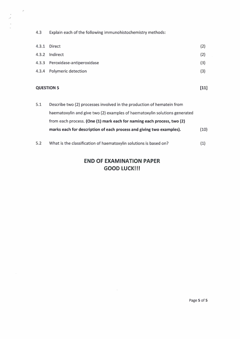
4.3 Explain each of the following immunohistochemistry methods:
4.3.1 Direct
(2)
4.3.2 Indirect
(2)
4.3.3 Peroxidase-antiperoxidase
(3)
4.3.4 Polymeric detection
(3)
QUESTION 5
[11]
5.1 Describe two (2) processes involved in the production of hematein from
haematoxylin and give two (2) examples of haematoxylin solutions generated
from each process. (One (1) mark each for naming each process, two (2)
marks each for description of each process and giving two examples).
(10)
5.2 What is the classification of haematoxylin solutions is based on?
(1)
END OF EXAMINATION PAPER
GOOD LUCK!!!
Page 5 of 5


