 |
ANP611S - ANATOMICAL PATHOLOGY 2A -1ST OPP - JUNE 2023 |
 |
1 Page 1 |
▲back to top |
nAmlBIA UnlVERSITY
OF SCIEnCE Ano TECHnOLOGY
FACULTYOF HEALTH, APPLIEDSCIENCESAND NATURAL RESOURCES
SCHOOL OF HEALTH SCIENCES
DEPARTMENT OF CLINICAL HEALTH SCIENCES
QUALIFICATION:BACHELOROF MEDICAL LABORATORYSCIENCES
QUALIFICATION CODE: 08BOHS
COURSECODE: ANP611S
LEVEL: 6
COURSENAME: ANATOMICAL PATHOLOGY2A
SESSION:
JUNE 2023
PAPER:
THEORY
DURATION: 3 HOURS
MARKS:
100
FIRST OPPORTUNITY EXAMINATION QUESTION PAPER
EXAMINER(S)
MS BELINDAROSELINTSAUSES
MODERATOR:
MS NDESHIPEWA HAMATUI-VALOMBOLA
INSTRUCTIONS
1. Answer ALL the questions.
2. Write clearly and neatly.
3. Number the answers clearly.
PERMISSIBLE MATERIALS
1. Non programmable calculator is allowed.
THIS QUESTION PAPERCONSISTSOF 5 PAGES(Including this front page)
 |
2 Page 2 |
▲back to top |
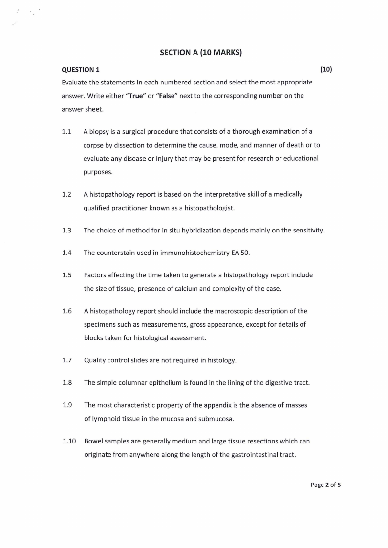
SECTION A (10 MARKS)
QUESTION 1
(10)
Evaluate the statements in each numbered section and select the most appropriate
answer. Write either "True" or "False" next to the corresponding number on the
answer sheet.
1.1 A biopsy is a surgical procedure that consists of a thorough examination of a
corpse by dissection to determine the cause, mode, and manner of death or to
evaluate any disease or injury that may be present for research or educational
purposes.
1.2 A histopathology report is based on the interpretative skill of a medically
qualified practitioner known as a histopathologist.
1.3 The choice of method for in situ hybridization depends mainly on the sensitivity.
1.4 The counterstain used in immunohistochemistry EA 50.
1.5 Factors affecting the time taken to generate a histopathology report include
the size of tissue, presence of calcium and complexity of the case.
1.6 A histopathology report should include the macroscopic description of the
specimens such as measurements, gross appearance, except for details of
blocks taken for histological assessment.
1.7 Quality control slides are not required in histology.
1.8 The simple columnar epithelium is found in the lining of the digestive tract.
1.9 The most characteristic property of the appendix is the absence of masses
of lymphoid tissue in the mucosa and submucosa.
1.10 Bowel samples are generally medium and large tissue resections which can
originate from anywhere along the length of the gastrointestinal tract.
Page2 of 5
 |
3 Page 3 |
▲back to top |
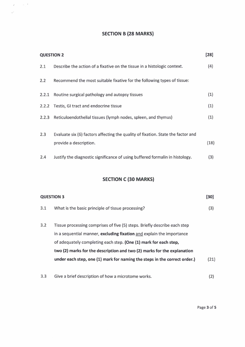
SECTION B (28 MARKS)
QUESTION 2
2.1 Describe the action of a fixative on the tissue in a histologic context.
2.2 Recommend the most suitable fixative for the following types of tissue:
2.2.1 Routine surgical pathology and autopsy tissues
2.2.2 Testis, GI tract and endocrine tissue
2.2.3 Reticuloendothelial tissues (lymph nodes, spleen, and thymus)
[28]
(4)
(1)
(1)
(1)
2.3 Evaluate six (6) factors affecting the quality of fixation. State the factor and
provide a description.
{18)
2.4 Justify the diagnostic significance of using buffered formalin in histology.
(3)
SECTION C (30 MARKS)
QUESTION 3
[30]
3.1 What is the basic principle of tissue processing?
(3)
3.2 Tissue processing comprises of five (5) steps. Briefly describe each step
in a sequential manner, excluding fixation and explain the importance
of adequately completing each step. (One (1) mark for each step,
two (2) marks for the description and two (2) marks for the explanation
under each step, one (1) mark for naming the steps in the correct order.)
(21)
3.3 Give a brief description of how a microtome works.
(2)
Page 3 of 5
 |
4 Page 4 |
▲back to top |
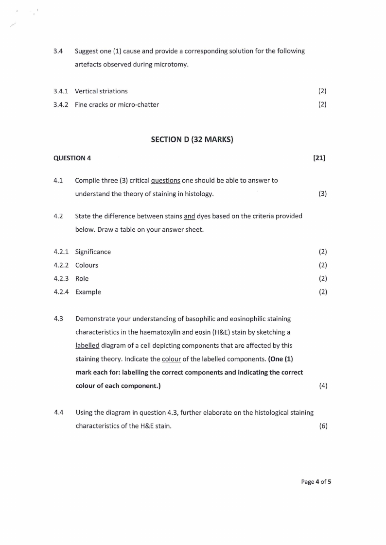
3.4 Suggest one {1) cause and provide a corresponding solution for the following
artefacts observed during microtomy.
3.4.1 Vertical striations
(2)
3.4.2 Fine cracks or micro-chatter
(2)
SECTION D (32 MARKS)
QUESTION 4
[21]
4.1 Compile three (3) critical questions one should be able to answer to
understand the theory of staining in histology.
(3)
4.2 State the difference between stains and dyes based on the criteria provided
below. Draw a table on your answer sheet.
4.2.1 Significance
(2)
4.2.2 Colours
(2)
4.2.3 Role
(2)
4.2.4 Example
(2)
4.3 Demonstrate your understanding of basophilic and eosinophilic staining
characteristics in the haematoxylin and eosin {H&E) stain by sketching a
labelled diagram of a cell depicting components that are affected by this
staining theory. Indicate the colour of the labelled components. (One (1)
mark each for: labelling the correct components and indicating the correct
colour of each component.)
(4)
4.4 Using the diagram in question 4.3, further elaborate on the histological staining
characteristics of the H&E stain.
{6)
Page 4 of 5
 |
5 Page 5 |
▲back to top |
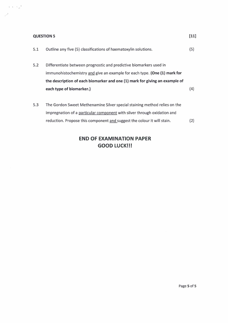
QUESTION 5
[11]
5.1 Outline any five (5) classifications of haematoxylin solutions.
(5)
5.2 Differentiate between prognostic and predictive biomarkers used in
immunohistochemistry and give an example for each type. (One (1) mark for
the description of each biomarker and one (1) mark for giving an example of
each type of biomarker.)
(4)
5.3 The Gordon Sweet Methenamine Silver special staining method relies on the
impregnation of a particular component with silver through oxidation and
reduction. Propose this component and suggest the colour it will stain.
(2)
END OF EXAMINATION PAPER
GOOD LUCK!!!
Page 5 of 5


