


 |
HAM621S - HAEMATOLOGY 2B - 2ND OPP- JAN 2023 |
|
|
1 Page 1 |
▲back to top |
nAmlBIA unlVERSITY
OF SCIEnCE Ano TECHnOLOGY
FACULTYOF HEALTH,APPLIEDSCIENCESAND NATURALRECOURCES
DEPARTMENT OF HEALTHSCIENCES
QUALIFICATION: BACHELOROF MEDICAL LABORATORYSCIENCES
QUALIFICATION CODE: 08BMLS
LEVEL: 6
COURSECODE: HAM621S
COURSENAME: HAEMATOLOGY 2B
SESSION:JANUARY 2023
DURATION: 3 HOURS
PAPER:THEORY
MARKS: 100
SUPPLEMENTARY/ SECONDOPPORTUNITYEXAMINATION PAPER
EXAMINER(S)
DR MAURICENYAMBUYA
MODERATOR:
DR ELZABEVAN DERCOLF
INSTRUCTIONS
1. Answer ALL the questions.
2. Write clearly and neatly.
3. Number the answers clearly.
PERMISSIBLEMATERIALS
1. Pen
2. Calculator
THIS QUESTION PAPERCONSISTSOF 8 PAGES(including this front page)
Page 1 of 8
|
|
2 Page 2 |
▲back to top |
SECTIONA [SO]
QUESTION 1
(20]
Select one correct answer to each questions below.
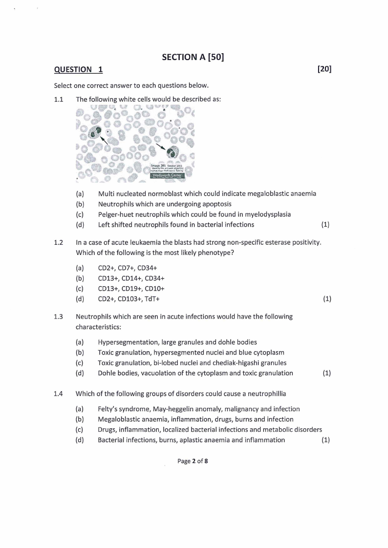
1.1 The following white cells would be described as:
]
• 381 OOot:,,:,'10
rtl't',rrowtdoti.!ltl,
·""""""'
(a) Multi nucleated normoblast which could indicate megaloblastic anaemia
(b) Neutrophils which are undergoing apoptosis
(c) Pelger-huet neutrophils which could be found in myelodysplasia
(d) Left shifted neutrophils found in bacterial infections
(1)
1.2 In a case of acute leukaemia the blasts had strong non-specific esterase positivity.
Which of the following is the most likely phenotype?
(a) CO2+,CD7+,CD34+
(b) CD13+,CD14+,CD34+
(c) CD13+,CD19+,CD10+
(d) CD2+,CD103+,TdT+
(1)
1.3 Neutrophils which are seen in acute infections would have the following
characteristics:
(a) Hypersegmentation, large granules and dohle bodies
(b) Toxic granulation, hypersegmented nuclei and blue cytoplasm
(c) Toxic granulation, bi-lobed nuclei and chediak-higashi granules
(d) Dahle bodies, vacuolation of the cytoplasm and toxic granulation
(1)
1.4 Which of the following groups of disorders could cause a neutrophillia
(a) Felty's syndrome, May-heggelin anomaly, malignancy and infection
(b) Megaloblastic anaemia, inflammation, drugs, burns and infection
(c) Drugs, inflammation, localized bacterial infections and metabolic disorders
(d) Bacterial infections, burns, aplastic anaemia and inflammation
(1)
Page 2 of 8
|
|
3 Page 3 |
▲back to top |
1.5 The following results were obtained on a sample of blood analysed in the
haematology laboratory. White cell count: 6.3 x 10⁹/1, Neutrophils: 28%, Band cells:
33%, metamyelocytes 8%, lymphocytes: 25%, Monocytes: 6 %. How would you
describe this blood picture?
(a) Leukaemoid reaction
(b) Left shift
(c) Leucoerythroblastic blood picture
(d) Granulocytosis
(1)
1.6 A hypercellular bone marrow with an M:E ratio of 1:10 indicates:
(a) Decreased erythropoiesis
(b) Increased erythropoiesis
(c) Increased myelopoiesis
(d) Decreased myelopoiesis
(1)
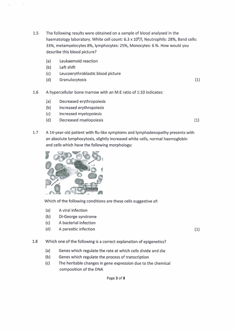
1.7 A 14-year-old patient with flu-like symptoms and lymphadenopathy presents with
an absolute lymphocytosis, slightly increased white cells, normal haemoglobin
and cells which have the following morphology:
Which of the following conditions are these cells suggestive of:
(a) A viral infection
(b) Di-George syndrome
(c) A bacterial infection
(d) A parasitic infection
(1)
1.8 Which one of the following is a correct explanation of epigenetics?
(a) Genes which regulate the rate at which cells divide and die
(b) Genes which regulate the process of transcription
(c) The heritable changes in gene expression due to the chemical
composition of the DNA
Page 3 of 8
|
|
4 Page 4 |
▲back to top |
(d) The inactivation of certain genes due to mutations which lead to
increased proliferation and decreased apoptosis
(1)
1.9 Which of the following described a chromosomal deletion?
(a) Point mutation resulting in a single aminoacid substitution
(b) Transfer of genetic material from one chromosome to another
(c) Loss of genetic material from a chromosome that does not appear on
another chromosome
(d) Duplication of a chromosome resulting in an increase of genetic material (1)
1.10 A patient has a platelet count of 1000 x 109/1.The platelets are abnormal in size,
shape and granularity. The white cell count is 12 x 109/1and the haemoglobin in
llg/dl. There is no Philadelphia chromosome. Which of the following is the most
likely diagnosis?
(a) Chronic Myeloid leukaemia
(b) Polycythaemia Vera
(c) Essential Thrombocythaemia
(d) Myelofibrosis
(1)
1.11 Acute leukaemias are often associated with which of the following?
(a) Bleeding
(b) Tiredness
(c) Bruising
(d) All of the above
(1)
1.12 The following surface marker results were obtained with lymphocytes from a 21-
year-old-man with a lymphocytosis of 16 x 109/L. Percentage of peripheral
lymphocytes reactive with antisera to: Kappa = 6%; Lambda = 4%; CD19 = 10%;
CD11c = 0%; T cells= 81%; CD56 (NK cells)= 6%. Which ONE is the most likely
diagnosis?
(a) Infectious mononucleosis
(b) Sq syndrome
(c) Chronic myelomonocytic leukaemia
(d) Sepsis
(1)
1.13 A 35-year-old man presented with weakness, lassitude and feeling easily tired. Her
bone marrow aspirate showed 15% myeloblasts and reduced erythropoiesis. What is
the most likely cause?
Page 4 of 8
|
|
5 Page 5 |
▲back to top |
(a) Acute myeloid leukaemia
(b) Acute lymphoid leukaemia
(c) Myelodysplastic syndrome
(d) Myelofibrosis
(1)
1.14 Dahle bodies are patches of dilated endoplasmic reticulum that appear as cerulean
blue cytoplasmic puddles and are mostly seen in_.
(a) Chronic myeloid leukaemia
(b) Leukemoid reaction
(c) Chediak higashi syndrome
(d) Infectious mononucleosis
(1)
1.15 A subtype M3 is associated with which of the following gene translocation.
(a) t(15;17)
(b) t(8;21)
(c) t(16;16)
(d) t(9;22)
(1)
Determine whether the following statements are True or False. Only select the correct letter
(a/b) corresponding to your answer.
1.16 Terminal deoxynucleotidyl transferase (Tdt) is a surface protein marker of mature B
cells.
(a) True
(b) False
(1)
1.17 Secondary immune deficiencies are rarely due to medicinal treatments.
(a) True
(b) False
(1)
1.18 Coulter's principle or cell impedance method uses fluorochromes to count cells.
(a) True
(b) False
(1)
1.19 In a leukemoid reaction, the NAP/LAP score is usually high.
(a) True
(b) False
(1)
Page 5 of 8
|
|
6 Page 6 |
▲back to top |
1.20 Lymphocytosis is characterised by lymphocyte count of >3.5 x10⁹/L.
{a) True
{b) False
{1)
QUESTION 2
[30]
A 28-year-old woman had a sore throat, fever and generalised lymphadenopathy. The
Doctor took blood samples and sent them to the laboratory for analysis and the initial
results were as follows.
White cell count
Haemoglobin
Platelets
14.0 X 10⁹/1
12.lg/dl
201 X 109/1
Neutrophils
39%
Lymphocytes
52%
Monocytes
5%
Band cells
2%
Eosinophils
2%
The granulocyte morphology was normal and acute leukaemia was ruled out. The
lymphocytes were however atypical and flow cytometry was requested
2.1 Calculate the absolute lymphocyte count and comment on the result.
(2)
2.2 What is the normal range for lymphocytes in both an adult female and 6 month
old baby
{2)
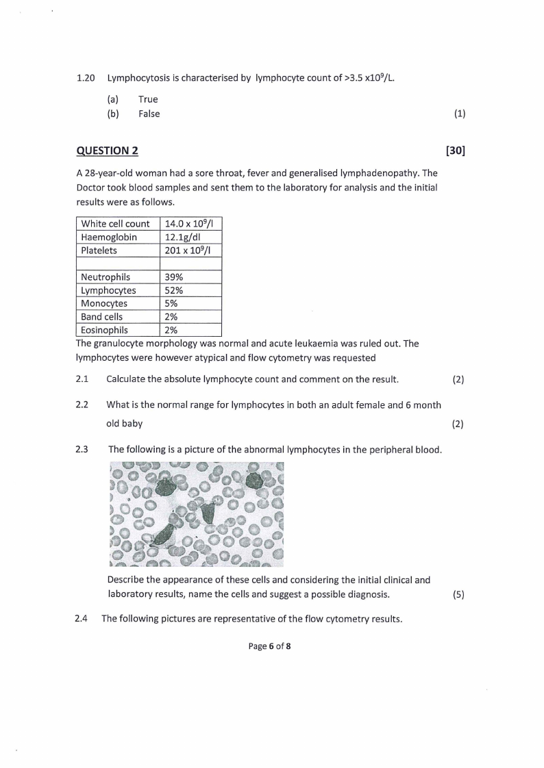
2.3 The following is a picture of the abnormal lymphocytes in the peripheral blood.
Describe the appearance of these cells and considering the initial clinical and
laboratory results, name the cells and suggest a possible diagnosis.
{S)
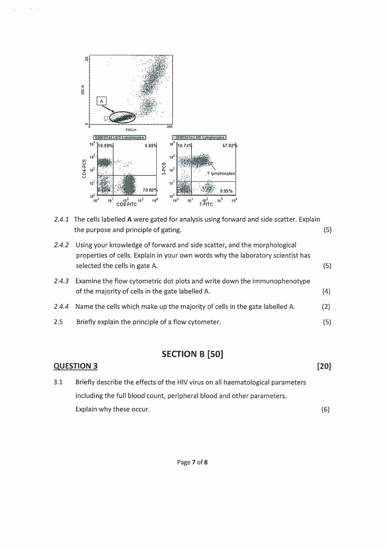
2.4 The following pictures are representative of the flow cytometry results.
Page6 of8
|
|
7 Page 7 |
▲back to top |
25S
FSC-H
0.99°
57.92~
10! • ·
\\:
"'q.(.:,
"' 102 ·
.
Cl
(.:,
cry
.. •T,_. _lymphocytes
101 ,
60°
10° -~:.;;:.;.,,1...-.:...iri~----!
10°
101
102
10;
CQS.flTC
101 -t't,-:~t---~----1.-:'.:~:iq~r:/J· I9J.9.9-%•.
102
7•FITC
2.4.1 The cells labelled A were gated for analysis using forward and side scatter. Explain
the purpose and principle of gating.
(5)
2.4.2 Using your knowledge of forward and side scatter, and the morphological
properties of cells. Explain in your own words why the laboratory scientist has
selected the cells in gate A.
(5)
2.4.3 Examine the flow cytometric dot plots and write down the immunophenotype
of the majority of cells in the gate labelled A.
(4)
2.4.4 Name the cells which make up the majority of cells in the gate labelled A.
(2)
2.5 Briefly explain the principle of a flow cytometer.
(5)
QUESTION 3
SECTIONB [SO]
3.1 Briefly describe the effects of the HIV virus on all haematological parameters
including the full blood count, peripheral blood and other parameters.
Explain why these occur.
[20]
(6)
Page 7 of 8
|
|
8 Page 8 |
▲back to top |
3.2 What peripheral blood morphological features should the Laboratory Scientist
look for in order to determine whether granulocytosis is due to a reactive or
infective process rather than a haemopoietic malignancy or leukaemia?
(5)
3.3 Explain the morphological differences between a blast and a promyelocyte.
(4)
3.4 Name two disorders in which Pseudo-Pelger Huet cells could be seen.
(2)
3.5 Name the three phases of carcinogenesis.
(3)
QUESTION 4
[30]
4.1 List 6 categories in which the World Health Organisation (WHO) classified
myelodysplastic syndrome into.
(6)
4.2 Of the MOS classification you have listed above, which category is most likely to
progress into acute myeloid leukaemia. Motivate your answer.
(4)
4.3 Discuss the pathogenesis, clinical findings as well as laboratory results of
chronic myelomonocytic leukaemia (CMML).
(12)
4.4 What disorder is CMML classified as according to the WHO? Explain the rationale
behind this classification.
(3)
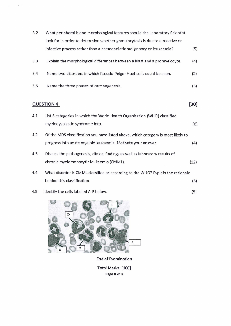
4.5 Identify the cells labeled A-E below.
(5)
A
End of Examination
Total Marks: [100]
Page8 of 8