ANP621S - ANATOMICAL PATHOLOGY - 1ST OPP - NOVEMBER 2024


ANP621S - ANATOMICAL PATHOLOGY - 1ST OPP - NOVEMBER 2024


1 Pages 1-10

▲back to top


1.1 Page 1

▲back to top


n Am I BI A u n IVER s I TY
OF SCIEnCE AnDTECHnOLOGY
Faculty of Health, Natural
Resources and Applied
Sciences
School of Health Sciences
Department of Clinical
Health Sciences
13 Jackson Kaujeua Street
Private Bag 13388
Windhoek
NAMIBIA
T: +264 61 207 2971
F: +264 61 207 997
E: dchs@nust.na
W: www.nust.na
QUALIFICATION : BACHELOR of MEDICAL LABORATORY SCIENCES
QUALIFICATION CODE: 08BMLS
LEVEL: 6
COURSE:ANATOMICAL PATHOLOGY
COURSECODE: ANP621S
DATE: NOVEMBER 2024
SESSION: 1
DURATION: 3 HOURS
MARKS: 100
FIRST OPPORTUNITY: QUESTION PAPER
EXAMINER:
MODERATOR:
Ms Belinda Roselin Tsauses
Ms Ndeshipewa Hamatui-Valombola
INSTRUCTIONS:
1. Answer all questions in the answer book.
2. Please write neatly and legibly.
3. Do not use the left side margin of the exam paper. This must be allowed for the
examiner.
4. No books, notes and other additional aids are allowed.
5. Mark all answers clearly with their respective question numbers.
PERMISSIBLE MATERIALS:
1. None.
ATTACHEMENTS
1. None.
This question paper consists of 6 pages including this front page.

1.2 Page 2

▲back to top


1.3 Page 3

▲back to top


SECTIONA: MULTIPLECHOICEAND TRUE FALSE
20 MARKS
QUESTION1: MULTIPLECHOICEQUESTIONS
[10 MARKS]
Evaluate the statements in each numbered section and select the most appropriate answer or
phrase from the given possibilities. Fill in the appropriate letter next to the number of the
correct statement/phrase in your ANSWERBOOK.
[10]
1.1 EA-36, EA-50, EA-65 and Orange G (OG-6) are examples of ________
a) Cytoplasmic stains.
b) Nuclear stains. H
c) H&E stains.
d) All of the above.
stains: (1)
1.2 Cytologists call the peppering effect of this infection on the surface of squamous epithelial
cells, which gives them a hazy blue appearance, as "clue cells".
(1)
a) Bacterial vaginosis.
b) Fungal elements.
c) Actinomyces.
d) Trichomonas vaginosis.
1.3 The organism possesses hair-like flagella, and on high magnification a smudgy grey
nucleus can be visualized with occasional tiny pink granules within the cytoplasm.
(1)
a) Bacterial vaginosis.
b) Leptothrix.
c) Actinomyces.
d) Trichomonas vaginosis.
1.4 Pseudohyphae of these organisms appears as eosinophilic tangled filaments with parallel
edges and without cross walls(septa) on the Pap stain.
(1)
a) Actinomyces.
b) Fungi.
c) Trichomonas vaginosis.
d) Leptothrix.
Anatomical Pathology (ANP621S)
1'1Opportunity November 2024
2

1.4 Page 4

▲back to top


1.5 Page 5

▲back to top


1.5 An anatomical distinct region of the female genital tract that is lined with
simple glandular epithelium, which forms branching crypts into the endocervical stroma. (1)
a) Endocervix.
b) Ectocervix.
c) Endocervical canal.
d) Squamocolumnar junction.
1.6 The opening of the cervical canal into the vaginal cavity is known as the:
(1)
a) Vaginal canal.
b) Endocervical canal.
c) Internal os.
d) External OS.
1.7 The opening of the cervical canal into the uterine cavity is known as the:
(1)
a) Endometrial canal.
b) Endocervical canal.
c) Internal os.
d) External OS.
1.8 The labia majora, labia minora, and clitoris are covered by:
(1)
a) Stratified squamous mucosa.
b) Keratinized stratified squamous epithelium.
c) Columnar mucinous epithelium.
d) Germinal epithelium.
1.9 The vagina is lined by:
(1)
a) Stratified squamous mucosa.
b) Keratinized stratified squamous epithelium.
c) Columnar mucinous epithelium.
d) Germinal epithelium.
Anatomical Pathology (ANP6215)
1st opportunity November 2024
3

1.6 Page 6

▲back to top


1.7 Page 7

▲back to top


(
(
1.10 The part of the female genital tract that consists of all the external parts and
tissues and includes the mans pubis, pudenda I cleft, labia majora, labia
minora, Bartholin's glands, clitoris and vaginal opening is known as the:
(1)
a) Uterus.
b) Cervix.
c) Vulva.
d) Vagina.
QUESTION 2: TRUE/FALSE QUESTIONS
[10 MARKS]
Evaluate the statements and select whether the statement is true or false. Write the
word 'True' or 'False' next to the corresponding number in your A_NSWERBOOK.
Correct each false statement by replacing the incorrect word(s) with the correct
word(s). (One (1) mark for stating either 'True' or 'False' and one (1) mark for
correcting each false statement.)
[10]
2.1 Most gynaecological specimens are vaginal smears, followed by smears of the cervix
or vulva.
2.2 Formalin is the preferred fixative for cytological specimens.
2.3 The H&E stain is the universal stain used for staining cytological samples.
2.4 If it is too thin, material may flake off in the staining process or result in cells
overlapping and therefore make interpretation difficult.
2.5 An artificial or morphological change in a cytological smear, which is visible by
light microscopy and is induced during sampling, transportation or smear preparation.
2.6 It is important to differentiate between air dried and fixed samples by appropriate
labelling to ensure that the correct staining method is applied.
Anatomical Pathology (ANP621S)
1st opportunity November 2024
4

1.8 Page 8

▲back to top


/

1.9 Page 9

▲back to top


Please answer ALL of the questions in this section.
[30 MARKS]
QUESTION 3:
[18 MARKS]
3.1 Explain the cytologic significance of the "transformation zone" of the cervix
and sketch a neatly labelled diagram depicting the transformation zone.
(Five (5) marks for the explanation and three (3) marks for the diagram.)
(8)
3.2 Describe five (5) factors that determine the type of epithelial cells seen in cervical
smears. Give your answers in point form.
(5)
3.3 Correlations between cytology and histology have identified two (2) features as
most useful for distinguishing normal and neoplastic cells. Briefly describe these
features.
(5)
QUESTION 4:
[12 MARKS]
4.1 Tabulate the difference between the three (3) phases of menopause according to
the following criteria:
4.1.1 Phases
(3)
4.1.2 Cytologic features
(6)
4.1.3 Maturation index (Ml)
(3)
SECTION C: LONG ANSWER QUESTIONS
Please answer ALL of the questions in this section.
[SO MARKS]
QUESTION 5:
[35 MARKS]
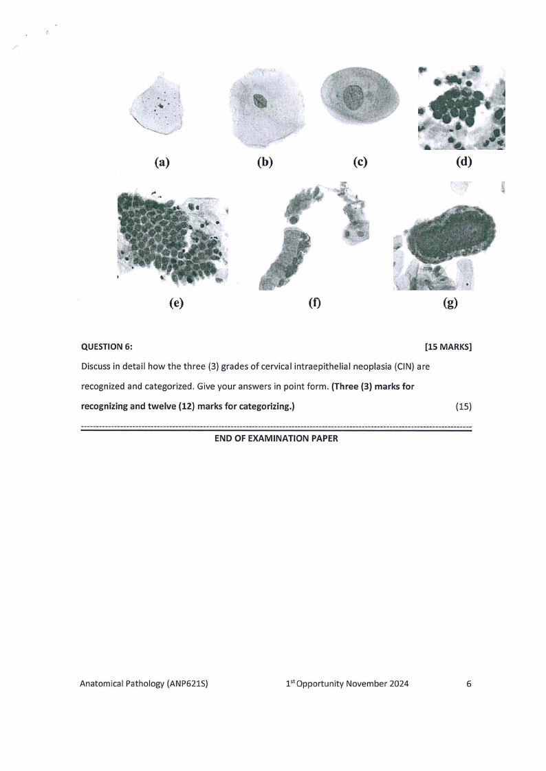
Identify and describe in detail the normal epithelial cells of the female genital tract
labelled from letter a - g below, based on their distinct cytological features observed
microscopically. Write the correct answer(s) next to the appropriate letter. PRINT IN
COLOUR. (One (1) mark for identification and four (4) marks for describing.)
(35)
Anatomical Pathology (ANP621S)
pt opportunity November 2024
5

1.10 Page 10

▲back to top


2 Pages 11-20

▲back to top


2.1 Page 11

▲back to top


(a)
(b)
(c)
(d)
(e)
(t)
(g)
QUESTION 6:
[15 MARKS]
Discuss in detail how the three (3) grades of cervical intraepithelial neoplasia (CIN) are
recognized and categorized. Give your answers in point form. (Three (3) marks for
recognizing and twelve (12) marks for categorizing.)
(15)
END OF EXAMINATION PAPER
Anatomical Pathology (ANP621S)
1'1Opportunity November 2024
6

2.2 Page 12

▲back to top


i'