


 |
ANP621S - ANATOMICAL PATHOLOGY - 2ND OPP - JANUARY 2025 |
|
|
1 Page 1 |
▲back to top |
nAmlBIA UnlVERSITY
OF SCIEnCE
Facultyof Health, Natural
Resourcesand Applied
Sciences
School of Health Sciences
Department of Clinical
Health Sciences
13Jackson Kaujeua Street
Private Bag 13388
Windhoek
NAMIBIA
T: +264 61 207 2970
F: +264 61 207 9970
E: dchs@nust.na
W: www.nust.na
QUALIFICATION: BACHELOR of MEDICAL LABORATORY SCIENCES
QUALIFICATIONCODE: 08BMLS
LEVEL:6
COURSE:ANATOMICAL PATHOLOGY
COURSECODE: ANP621S
DATE: JANUARY 2025
SESSION: 1
DURATION: 3 HOURS
MARKS: 100
SECOND OPPORTUNITY/ SUPPLEMENTARY: EXAMINATION QUESTION PAPER
EXAMINER:
MODERATOR:
Ms Belinda Roselin Tsauses
Ms Ndeshipewa Hamatui-Valombola
INSTRUCTIONS:
1. Answer all questions in the answer book.
2. Please write neatly and legibly.
3. Do not use the left side margin of the exam paper. This must be allowed for the
examiner.
4. No books, notes and other additional aids are allowed.
5. Mark all answers clearly with their respective question numbers.
PERMISSIBLE MATERIALS:
1. None.
ATTACHMENTS
1. None.
This question paper consists of 7 pages including this front page
|
|
2 Page 2 |
▲back to top |
~SECTIONA: MULllPLE CHOICE AND TRUE/ FALSE
[20 MARKS]
QUESTION 1: MULTIPLE CHOICE QUESTIONS
[10 MARKS]
Evaluate the statements in each numbered section and select the most appropriate answer
or phrase from the given possibilities. Fill in the appropriate letter next to the number of the
correct statement/phrase in your ANSWERBOOK.
[10]
1.1 The ____
is the neck of the uterus, the lower, narrow portion where it joins with
the upper part of the vagina.
(1)
a) Internal os.
b) External OS.
c) Cervix.
d) Endometrium.
1.2 A _______
_, also known as the germinal epithelium, surrounds the ovary. (1)
a) Mesothelium.
b) Stratified squamous mucosa.
c) Columnar mucinous epithelium.
d) None of the above.
1.3 In the _____
__, the stroma is composed of many small fibroblastic cells along
with scattered lymphocytes, macrophages, and blood vessels.
(1)
a) Vagina.
b) Fallopian tubes.
c) Ovaries.
d) Uterus.
1.4 Epithelial cells can be described by their _____
and ______
_
(1)
a) Structure; function.
b) Morphology; staining properties.
c) Nucleus; cytoplasm.
d) None of the above.
Anatomical Pathology (ANP621S)
2
2nd Opportunity- January2025
|
|
3 Page 3 |
▲back to top |
1.5 The term ______
is used to describe a finely granular chromatin pattern.
(1)
a) Vesicular.
b) Pyknotic.
c) Bland.
d) None of the above.
1.6 In Pap staining, the intensity of nuclear staining can be described as:
(1)
a) Hyperchromatic.
b) Normochromatic.
c) Hypochromatic.
d) All of the above.
1.7 In cervical smears or Liquid Based Cytology (LBC)slides, _______
_
generally present as discrete single cells.
(1)
a) Endometrial cells.
b) Intermediate squamous cells.
c) Superficial squamous cells.
d) Endocervical cells.
1.8 What type of cytology sample is used during cytocentrifugation?
(1)
a) Biopsy.
b) Conventional smear.
c) LBC.
d) None of the above.
1.9 These tumors do not necessarily turn into malignant tumors:
(1)
a) Precancerous lesions.
b) Carcinoma in - situ.
c) Benign.
d) None of the above.
Anatomical Pathology (ANP621S)
3
2nd Opportunity- January2025
|
|
4 Page 4 |
▲back to top |
/.
1.10 Reparative changes can be atypical and mimic---------~
because when undergoing repair cells are depleted of normal glycogen
content
may have nuclear atypia.
(1)
a) Pre-cancerous lesions.
b) Cancerous lesions.
c) Benign changes.
d) Carcinoma in - situ.
QUESTION 2: TRUE/FALSE QUESTIONS
[10 MARKS]
Evaluate the statements and select whether the statement is true or false. Write the
word 'True' or 'False' next to the corresponding number in your ANSWERBOOK.
Correct each false statement by only replacing the incorrect word(s) with the correct
word(s).
[10]
2.1 EndoPaps refer to the process of collection by inserting a brush through the
endocervical canal and sampling areas of the cervix.
2.2 The microscope concentrates small numbers of cells suspended in fluids into a
6mm diameter cycle onto a glass slide for subsequent microscopic observation
2.3 The amount of material aspirated from a fine needle aspiration biopsy specimen is so
small that it is almost impossible to fix the smear before air drying occurs.
2.4 EA-65 is a cytoplasmic stain that helps to differentiate adenocarcinomas of the
endocervix (pink) from those of endometrium (blue).
2.5 In wet fixation, the cells should be exposed to air and the slide should remain
submerged for a minimum of 15-30 minutes.
2.6 There are four (4) categories of fixation methods that are used in the cytology
laboratory.
Anatomical Pathology (ANP6215)
4
2nd Opportunity- January2025
|
|
5 Page 5 |
▲back to top |
SECTION B: SHORT ANSWER QUESTIONS
Pleaseanswer ALL of the questions in this section.
QUESTION 3:
3.1 Name and describe four (4) different types of artefacts commonly seen in
Cytology, including the cause. Present your answers in a table.
[33 MARKS]
[17 MARKS]
(12)
3.2 Give a general cytological description of metaplastic cells.
(5)
QUESTION 4:
4.1 Briefly explain the significance of progesterone levels in the presence of
navicular cells and common conditions in which they may be seen.
[16 MARKS]
(4)
4.2 To justify the importance of intermediate cells undergoing cytolysis, explain the
process of cytolysis from a cytologic perspective and describe how these cells will
look like on an LBCslide. Give your answers in point form. (Seven (7) marks for
explaining and one (1) mark for describing.)
(8)
4.3 With reference to your answer(s) in question 4.2, state the conditions under
which the process of cytolysis will not take place. Give reason(s) for your
answer(s).
(4)
Anatomical Pathology (ANP621S)
5
2nd Opportunity- January 2025
|
|
6 Page 6 |
▲back to top |
t'
SEc:TION C: tONG ANSWER QUESTIONS
Please answer ALL of the questions in this section.
[47 MARKS]
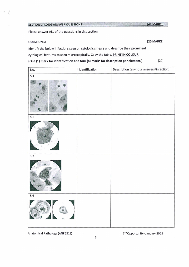
QUESTION 5:
[20 MARKS]
Identify the below infections seen on cytologic smears and describe their prominent
cytological features as seen microscopically. Copy the table. PRINT IN COLOUR.
(One (1) mark for identification and four (4) marks for description per element.)
(20)
No.
Identification
Description (any four answers/infection)
5.1
5.2
5.3
5.4
Anatomical Pathology (ANP6215)
6
2nd Opportunity- January 2025
|
|
7 Page 7 |
▲back to top |
r
I
QUESTION 6:
[27 MARKS]
6.1 Sketch a scenario to explain the process of metastasis in detail, using breast
cancer as an example. Give your answers in point form.
(15)
6.2 Discuss the concept of invasion of cancer cells.
(12)
END OF QUESTION PAPER.
Anatomical Pathology (ANP6215)
7
2nd Opportunity- January 2025